杂志信息/Information

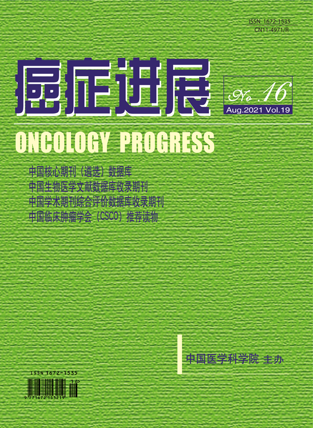
- 刊名:癌症进展
- Oncology Progress Journal
- 主管:国家卫生健康委员会
- 主办:中国医学科学院
- 社长:张凌
- 主编:赵平
- 编辑部主任:穆红
- 出版单位:中国协和医科大学出版社有限公司
《癌症进展》编辑部
100730,北京东单三条9号
联系电话:010-57528109
E-mail:azjzzz@163.com
http://www.aizhengjinzhan.com - 印刷:北京联合互通彩色印刷有限公司
- 国内统一连续出版物号:CN 11-4971/R
- 国际标准连续出版物号ISSN 1672-1535
下载专区/Download
订阅电子期刊/Subscribe
提交您的邮箱地址,我们会定期将电子期刊 发送到您的邮箱
期刊检索/Journal Search
扫一扫,关注

2016 年第 1 期 第 14 卷
食管癌转移淋巴结CT诊断标准探讨
作者:
单位:
- 摘要:
- 【摘要】摘要:目的 通过薄层CT多平面重建图像所见与病理结果对照,探讨转移淋巴结诊断标准。方法 以病理为金标准,回顾分析208例胸段食管癌患者淋巴结的术前多层螺旋CT多平面重建图像, 测量淋巴结的长径、短径,计算出直径比(短径/长径,将淋巴结直径比≥0.7视为圆形淋巴结),同时观察淋巴结有无环形强化,成簇分布、边缘模糊等影像学特征,分析比较各种指标的诊断价值。结果 以短径≥1.0cm作为转移淋巴结的诊断标准,敏感度为23.6%,特异度为98.6%,约登指数为0.22;以短径≥0.8cm作为转移淋巴结的诊断标准,敏感度为47.6%,特异度为93.9%,约登指数为0.42;以短径≥0.6cm作为转移淋巴结的诊断标准,敏感度为83.8%,特异度为65.8%,约登指数为0.50。以短径≥0.8cm作为转移淋巴结诊断标准,短径<0.8cm的淋巴结运用直径比≥0.7作为诊断转移的标准,敏感度为83.8%,特异度为82.9%,约登指数为0.67。以环形强化或边缘模糊征象作为诊断标准的阳性预测值分别为62.2%、89.4%。结论 用较小的短径作为转移淋巴结的诊断标准能提高诊断效能,直径比有助于提高CT术前诊断转移淋巴结的价值,边缘模糊、环形强化可作为转移淋巴结的参考诊断指标。
- Abstract: Objective To study the morphological criteria of lymph node metastasis in esophageal cancer. Method The MSCT MPR of 208 cases of esophageal cancer that were confirmed pathologically were characterized retrospectively. Lymph node were considered round if the axial ratio exceeded 0.7.Internal lymph node structures were also evaluated. Result When metastatic lymph nodes are de?ned as those with a short-axis dimension greater than 1.0 cm、0.8cm、0.6cm respectively,the sensitivity were 23.6%、47.6% and 83.8% respectively,and specificity were 98.6% 、93.9%and 65.8% respectively; When metastatic lymph nodes are de?ned as those with a dimension greater than 0.6cm,the sensitivity and specificity were 83.8% and 65.8%; When metastatic lymph nodes are de?ned as those with a short-axis dimension greater than 0.8cm and those with a short-axis diameter shorter than 0.8cm in terms of with axial ratio ( short diameter /major diameter) exceeded 0.7,the sensitivity and specificity were 83.8% and 82.9%.The positive predictive value(PPV) were 62.2% and 89.4% based on lymph nodes metastasis with obscure or rim respectively. Conclusion The value of CT diagnosis of esophageal cancer lymph node metastasis can be improved if with shorter axis dimension criteria ,and diameter ratio is also helpful to improve the value of CT preoperative diagnosis of metastasis lymph nodes, edge blur, rin enhancement can be additional criterias for diagnosis of lymph nodes metastasis.


